Marie and Loni's project
August 12, 2010
qPCR : Primers test on Phase 1
Samples from the same treatment were pooled : 1.5 µl of each individual/4 treatments (H (Heat), V (VT), HV (Heat and VT), C (control)).
5 new pair of primers : FoxO, Interleukin, Toll, NF-kß, TRAF6
-> 4 samples (4 "treatments") + 2 negative controls /set of primers
Master mix for each pair of primers (Volume t : 25 µl)
- 2X SyBr Green MM : 12.5 µl
- BSA : 1.5 µl
- Forward primer : 0.5 µl
- Reverse primer : 0.5 µl
- sterile H20 : 9 µl
- 1 µl of pooled cDNA
qPCR plate map :
| 1 |
2 |
3 |
4 |
|
| A |
H_IL |
VH_Fox |
Neg_NF |
H_T6 |
| B |
V_IL |
C_Fox |
Neg_NF |
V_T6 |
| C |
VH_IL |
Neg_Fox |
HV_Toll |
VH_T6 |
| D |
C_IL |
Neg_Fox |
V_Toll |
C_T6 |
| E |
Neg_IL |
H_NF |
H_Toll |
Neg_T6 |
| F |
Neg_IL |
V_NF |
Neg_Toll |
Neg_T6 |
| G |
H_Fox |
VH_NF |
C_Toll |
|
| H |
V_Fox |
C_NF |
Neg_Toll |
FoxO : 55.1°C / 54.3°C
Toll : 63.7°C / 62.8°C
TRAF-6 : 62.0°C / 62.2°C
IL : 63.9°C / 63.2°C
NF : 62.7°C / 61.8°C
Annealing temperature for the qPCR = 54°C
August 11, 2010
Western Blot : no magic :-(
H1, H2, C1, C2, V1, V2, HV1, HV2, Barnacle heat stress ON (positive control)
25µl of extracted proteins + 25 µl of 2X Reducing Sample Buffer
Run on a Precise Protein Gel : 150V-45mins
Transfer on the membrane : 30 min-20V
NB: Primary antibody solution = Blocking Solution 10 ml + HSP 70 antibody 9.9 µl
Incubation with primary antibody 1h45
Revelation : no magic.... Even for the barnacle sample...
The gel was stained with Comassie Stain and we could see that a lot of proteins was loaded on the gel... Problem during the transfer on the membrane ? Problem during the western ? No binding of this antibody ?
Vt plates for counting
To assess the Vt concentration inoculated during the experiment, we streaked tenfold serial dilutions on TCBS agar.
RNA extraction, Phase 2-1
H1-8, V1-8
~50-100mg of tissue stored at -80°C
Following the Trizol protocol
The RNA pellet was resuspended in 50 µl of DEPC-water (very small pellets after centrifugation)
Storage at -80°C.
August 10, 2010
Phase 2, part 2: VT stress and heat and VT stress experiments
to begin, the VT cultures we grew overnight were centrifuged (6, 50 ml tubes) at 4500g with an acceleration of 6
the VT pellets were resuspended in 8 mls of 0.22um FIltered Sea Water and the samples were pooled
animals were photographed in the sea table befor ethe experiment
each animal was inoculated with 2 mls of the VT culture
VT= 2 mls Vt culture each at room temperature
VTH= 2 mls Vt culture each at 35C in the heat bath
Vt dilutions were made and 100ul was spread onto Marine Agar plates 10^1 - 10^5
the cfu's will be counted tmr. so we can estimate the amount of Vt each anemone was exposed to
for the stress experiments the animals were photographed at= 9:00am, 10:00am, 11:00am, 12:00pm
after the stress the animals were weighed, dissected and the tentacles were collected and stored for RNA and protein extraction
Weight (g):
| V1 |
3.38 |
HV1 |
9.74 |
C5 |
1.88 |
||
| V2 |
1.55 |
HV2 |
1.21 |
C6 |
0.71 |
||
| V3 |
1.52 |
HV3 |
1.57 |
C7 |
0.67 |
||
| V4 |
4.68 |
HV4 |
1.53 |
C8 |
1.36 |
||
| V5 |
1.18 |
HV5 |
2.83 |
||||
| V6 |
2.26 |
HV6 |
1.52 |
||||
| V7 |
4.21 |
HV7 |
2.74 |
||||
| V8 |
2.95 |
HV8 |
1.14 |
notes:
V: partially/ attached some mucus produced, contracting body and tentacles
HV: not attached, some minor body contraction, no tentacle contraction
C: minor contractions of the body and tentacles, some mucus produces during dissection
qPCR - ßactin
Run of Phase 1.5 samples to normalize results from 8/9.
Master mix for each pair of primers (Volume t : 25 µl)
- 2X SyBr Green MM : 12.5 µl
- BSA : 1.5 µl
- Forward primer : 0.5 µl
- Reverse primer : 0.5 µl
- sterile H20 : 9 µl
- 1 µl of cDNA
Amplification worked without any dimers detection. Contamination in 1 of the 2 negative controls (CT=38.85)...
Ct values and Arbitrary expression values (AEV=10^(-(0.3012 *CT) + 11.434))
| HV1 |
HV2 |
HV3 |
HV4 |
H1 |
H2 |
H3 |
H4 |
V1 |
V2 |
V3 |
V4 |
C1 |
C2 |
C3 |
|
| CT |
29.42 |
26.63 |
27.2 |
27.56 |
30.51 |
28.5 |
26.39 |
26.9 |
27.48 |
31.89 |
27.46 |
28.05 |
25.83 |
ND |
29.12 |
| AEV |
373.8 |
2588 |
1743 |
1358 |
175.5 |
707.6 |
3057 |
2146 |
1436 |
67.41 |
1456 |
966.8 |
4508 |
ND |
460.4 |
Extraction of the 32 samples from Phase 2 stored at -80°C
~25mg of tissue homogenized in 500µl of CellLytic MT Solution
Spin 10min-12000g-4°C
Save supernatant -> stored at -20°C
August 9, 2010
Phase 1.5 : qPCR
Test of all individual samples from Phase I with SOD primers to assess the interindividual variability.
Same master mix recipe as in Phase I (no sign of dimers fluorescence).
Master mix for each pair of primers (Volume t : 25 µl)
- 2X SyBr Green MM : 12.5 µl
- BSA : 1.5 µl
- Forward primer : 0.5 µl
- Reverse primer : 0.5 µl
- sterile H20 : 9 µl
- 1 µl of cDNA
qPCR plate map :
| 1 |
2 |
3 |
|
| A |
HV1 |
V1 |
Neg |
| B |
HV2 |
V2 |
|
| C |
HV3 |
V3 |
|
| D |
HV4 |
V4 |
|
| E |
H1 |
C1 |
|
| F |
H2 |
C2 |
|
| G |
H3 |
C3 |
|
| H |
H4 |
Neg |
qPCR results analysis
Amplification worked without any dimers detection and contamination in negative controls!
Ct values and Arbitrary expression values (AEV=10^(-(0.3012 *CT) + 11.434))
| HV1 |
HV2 |
HV3 |
HV4 |
H1 |
H2 |
H3 |
H4 |
V1 |
V2 |
V3 |
V4 |
C1 |
C2 |
C3 |
|
| CT |
36.38 |
32.07 |
34.8 |
36.73 |
39 |
37.08 |
32.09 |
32.49 |
34.46 |
ND |
36.23 |
ND |
39 |
37.08 |
32.09 |
| AEV |
2.995 |
59.5 |
8.959 |
2.349 |
0.487 |
1.843 |
58.68 |
44.46 |
11.34 |
ND |
3.323 |
ND |
5.747 |
0.473 |
7.326 |
Heat Stress Experiment
At 8:05AM the heat bath was turned on and set for 35C
8:10AM
we made VT cultures to be used tmr. by adding VT colonies to 50 mls of Marine Broth (in 5 tubes) and let to grow overnight on the belly dancer in lab 10
8:25AM
the temperature in the sea table was measured at 12C
the temperature in the hot bath reached 35C
anemones were photographed in the sea table before they were transferred to the hot bath
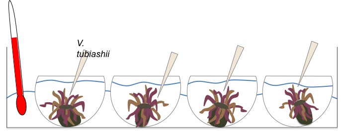
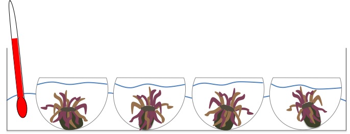
Anemones H1-H8 were removed from the sea table, the glass dishes were wiped down to avoid getting seawater in the heat bath and then moved to the heat bath
An extra dish was placed in the heat bath with just seawater and a thermometer
this way we could monitor the temperature of the heat bath and the temperature of the water that the anemones were subjected to in the dishes
the anemones were photographed at the begining of the experiment and every hour until the end= 8:30, 9:30, 10:30, 11:30
at 11:30AM the anemones were weighed, dissected and the tentacles were collected into two tubes for each animal: 1 for RNA work and 1 for protein work
Weights (g):
| H1 |
3.33 |
C1 |
8.01 |
|
| H2 |
3.00 |
C2 |
1.93 |
|
| H3 |
4.44 |
C3 |
6.35 |
|
| H4 |
3.03 |
C4 |
3.71 |
|
| H5 |
2.59 |
|||
| H6 |
6.81 |
|||
| H7 |
3.60 |
|||
| H8 |
2.09 |
heat stressed animals were partially or not attached to dishes upon removal for weight measurements and not very responsive, animals did not retract tentacles during dissection
control animals were contracting normally: body and tentacles, making it difficult to find and remove tentacles
tentacle samples were put in tubes for protein and RNA extraction and put on dry ice until stored at -80C
August 8, 2010
Anemone Collection (Phase 2)
Anemones were collected from the intertidal at Cattle Point (poo site) at 10AM.
35 animals were collected in a plastic bag with water and were brought bag to lab 5 in a cooler.
The anemones were weighed and placed in labeled, glass dishes in the sea table (2 anemones per dish).
8 animals will be used per treatment : Heat (H), VT (V), Heat and VT (HV), and Control (C)
Animals and weight(g) :
| H1 |
3.41 |
V1 |
2.25 |
HV1 |
7.09 |
C1 |
3.60 |
|||
| H2 |
2.46 |
V2 |
0.76 |
HV2 |
1.40 |
C2 |
3.60 |
|||
| H3 |
2.40 |
V3 |
1.21 |
HV3 |
1.56 |
C3 |
5.30 |
|||
| H4 |
2.15 |
V4 |
1.19 |
HV4 |
1.65 |
C4 |
1.44 |
|||
| H5 |
1.72 |
V5 |
1.40 |
HV5 |
3.29 |
C5 |
1.82 |
|||
| H6 |
2.56 |
V6 |
1.76 |
HV6 |
1.23 |
C6 |
1.02 |
|||
| H7 |
2.56 |
V7 |
3.93 |
HV7 |
1.57 |
C7 |
1.13 |
|||
| H8 |
1.46 |
V8 |
2.37 |
HV8 |
1.87 |
C8 |
0.85 |
August 7, 2010
QPCR results analysis (of Phase 1)
We got a strong amplification for VWR and ßactin, low amplification for SOD and S1PP2 and no amplification for PSAP.
All of the negative controls dissociation curves were flat, except for VWR and ßactin but the peak corresponded to dimers. We're going to need to improve the primer concentrations for those 2 primers.
-> no contamination !
All of the sample dissociation curves showed aligned peaks for the 4 treatments (H, V, HV and C) with different fluorescence intensities.
Ct values (except for the negative controls) were used to calculate the arbitrary expression value for each (pooled) sample using the following equation : AEV=10^(-(0.3012 *CT) + 11.434)
ßactin was chosen to be our reference gene.
Data were then normalized by dividing the AEV of the gene of interest by the AEV of ßactin (reference gene).
| VWF |
SOD |
ßactin |
S1PP2 |
|||||||||||||||||
| V |
H |
VH |
C |
V |
H |
VH |
C |
V |
H |
VH |
C |
V |
H |
VH |
C |
V |
H |
VH |
C |
|
| CT |
26.98 |
26.36 |
26.50 |
24.41 |
33.9 |
30.82 |
31.94 |
35.03 |
20.92 |
21.55 |
21.69 |
21.24 |
35.27 |
31.45 |
ND |
29.58 |
39.65 |
38.92 |
37.28 |
34.3 |
| AEV |
2030.6 |
3121.53 |
2832.7 |
12070.02 |
16.72 |
141.59 |
65.11 |
7.64 |
135798.82 |
87728.36 |
79610.8 |
108770.72 |
6.47 |
91.47 |
ND |
334.58 |
0.31 |
0.51 |
1.6 |
12.62 |
--> Phase 1.5 (for Monday): Run a QPCR of every single sample (not pooled by treatment) using SOD primers (15 samples + 2 negative controls) to see there is a significant difference in the expression of the amplified gene (investigate individual variation).
August 6, 2010
RNA extraction (Phase 1 continued)
Following the manufacturer's instructions, RNA was extracted from all of the 15 samples (50-100mg of tissues sampled for proteinextraction and stored at -80°C were previously weighed !).
The RNA pellet was resuspended in 100 µl of DEPC-water
Storage at -80°C.
Reverse transcription
- 17.5 µl of RNA was heated at 70°C for 5min (plate in the thermocycler)
- RNA was transfered to ice for 5-10 min
- 7.5 µl of Master Mix containing :
5 µl MMLV Buffer 5X
1.25 µl dNTPs 10mM
0.5 µl MMLV RTase
0.5 µl oligo dT primer
were added and the plate was set in the thermocycler at 37°C for 1h
and then at 95°C for 3min
The cDNA was stored on ice in the freezer (-20C) until the qPCR
qPCR
To test the primers available, samples from the same treatment were pooled : 2 µl of each individual/4 treatments (H (Heat), V (VT), HV (Heat and VT), C (control)).
We tested the 5 pairs of primers available in the lab and designed for A. elegantissima (S. Roberts) : VWR, b actin, SOD, S1PP2, PSAP
-> 4 samples (4 "treatments") + 2 negative controls /set of primers
Master mix for each pair of primers (Volume t : 25 µl)
- 2X SyBr Green MM : 12.5 µl
- BSA : 1.5 µl
- Forward primer : 0.5 µl
- Reverse primer : 0.5 µl
- sterile H20 : 9 µl
- 1 µl of pooled cDNA
qPCR plate map :
| 1 |
2 |
3 |
4 |
|
| A |
V_VWF |
VH_SOD |
Neg_Bactin |
V_PSAP |
| B |
H_VWF |
C_SOD |
Neg_Bactin |
H_PSAP |
| C |
VH_VWF |
Neg_SOD |
V_S1PP2 |
VH_PSAP |
| D |
C_VWF |
Neg_SOD |
H_S1PP2 |
C_PSAP |
| E |
Neg_VWF |
V_Bactin |
VH_S1PP2 |
Neg_PSAP |
| F |
Neg_VWF |
H_Bactin |
C_S1PP2 |
Neg_PSAP |
| G |
V_SOD |
VH_Bactin |
Neg_S1PP2 |
|
| H |
H_SOD |
C_Bactin |
Neg_S1PP2 |
VWF-F : 55.1°C / VWF-R : 57.1°C
S1PP2-F : 55.3°C / S1PP2-R : 52.8°C
SOD-F : 54.3°C / SOD-R : 53.0°C
ßactin-F : 56.6°C / -R : 57.3°C
PSAP-F : 53.8°C / PSAP-R : 52.8°C
Annealing temperature for the qPCR = 55°C
qPCR program :

August 5, 2010
Vt inoculation / Vt+Heat stress experiments (continuing in Phase 1)


Preparation of the "Vt solution" to inoculate the individuals : 5 ml of Vt culture (marine broth) was pellet
ed. The pellet was resuspend in 250 µl of sterile seawater (0.22um FSW).
Hot bath set for 35°C
t0 : Vt was added just above the anemones oral disk with a pipette
Pictures of each individual were taken every 15min.
Each individual was weighed, cut in half and tentacles were removed to be sampled for further RNA and protein extraction and stored at
-80°C.
RNA extraction
RNA extraction of all 15 samples (4 indivuals/3 treatments + 1 control/treatment) following the Trizol protocol.
We forgot to weigh the amount of tissue extracted, so the extraction failed !
The "half-extracted" samples were stored at -80°C just in case !
August 4, 2010
Preliminary experiment: Phase 1
Samples collected at Cattle point on 7/31.
Preparation for trial runs:
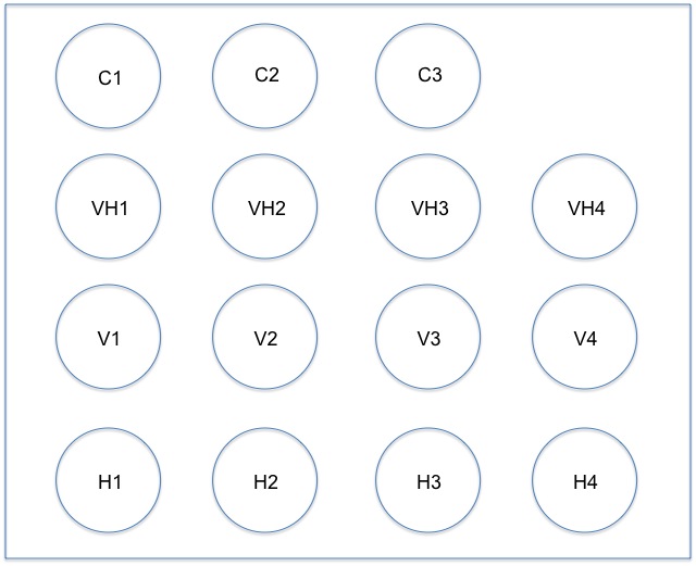
Loni and Marie separated anemones into individual (labeled) glass dishes in the sea table to acclimate
We made a map of the position of the dishes on the outside of the tank

The temperature of the water in the sea table is 12-15C
All 13 anemones were weighed and divided into test groups as follows:
Heat 35C
H1 4.57g
H2 3.12g
H3 4.37g
H4 4.42g
Vibrio tubiashii
V1 1.88g
V2 3.10g
V3 0.68g
V4 3.44g
Vibrio tubiashii and Heat 35C
VH1 4.22g
VH2 4.19g
VH3 3.29g
VH4 2.81g
Control (for our test runs we only have 1 animal per treatment)
C1 1.38g
C2 2.97g
C3 1.38g
We set up VT serial dilutions: 10^1, 10^2, 10^3, 10^4, 10^5, 10^6, 10^7, 10^8, 10^9, 10^10
by adding 4.5 mLs of dIH2O to 10 falcon tubes and then adding 0.5 mLs of our VT culture to the first dilution, mixing well, and then adding 0.5 mLs from that tube to the next, mixing well and continuing on...
We plated these dilutions along with the pure VT culture and the dIH20 that was used for the dilutions
We set up a new culture today by adding 49 mLs of marine broth to a 50 mL tube and addind 1 mL of our inital VT culture to that tube, O/N on the belly dancer in lab 10
The growth on these plates will give us an idea of ~how many VT we grow over night
When we do our real experiment we will probably only plate the concentration that we use to expose the anemones to
The hot water bath was set for 35C
Heat stress trial run:
The hot water bath was ready to go in the afternoon at 35C
The experimental individuals were photographed in the sea table in their happy state
We took the 4 selected individuals (H1-H4) out of the sea table and wiped down the outside of the glass dishes to not get any sea water into the heat bath

Heat stress: 3h
Photographs taken every 15min and temperature checked
At the end of the stress, individuals (4 + control) were weighed and immediately dissected (first we cut longitudinally then we cut horizontally to remove the tentacles.
Half of the tentacles were stored for proteins,the other half for RNA (-80°C).